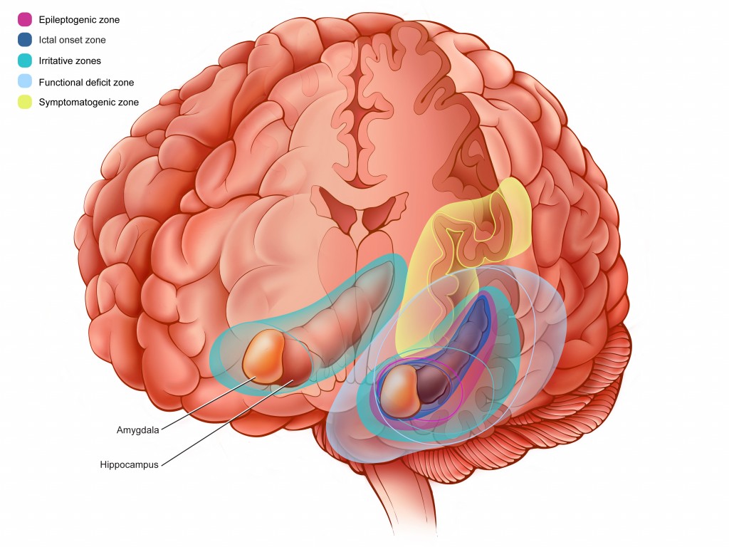
Illustration showing an epileptic network. These are regions of the brain that may be involved during reoccurring epileptic seizures.
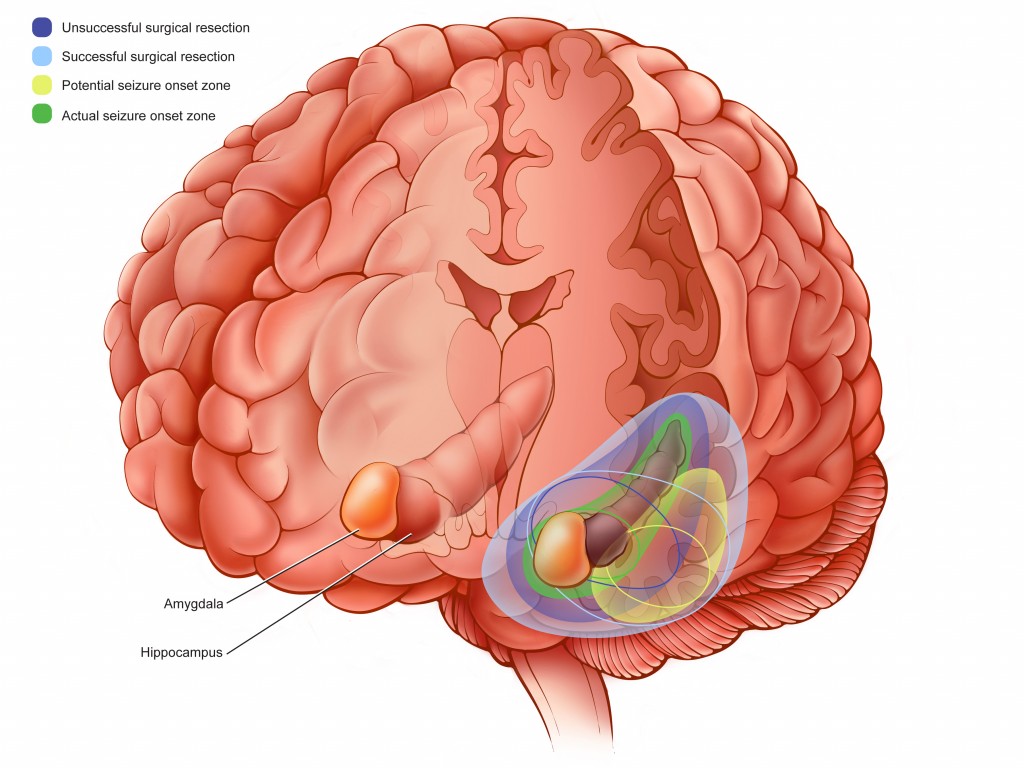
An illustration showing surgical options for patients with reoccurring epileptic seizures. Focal resection surgery removes a portion of the brain responsible for the seizures.
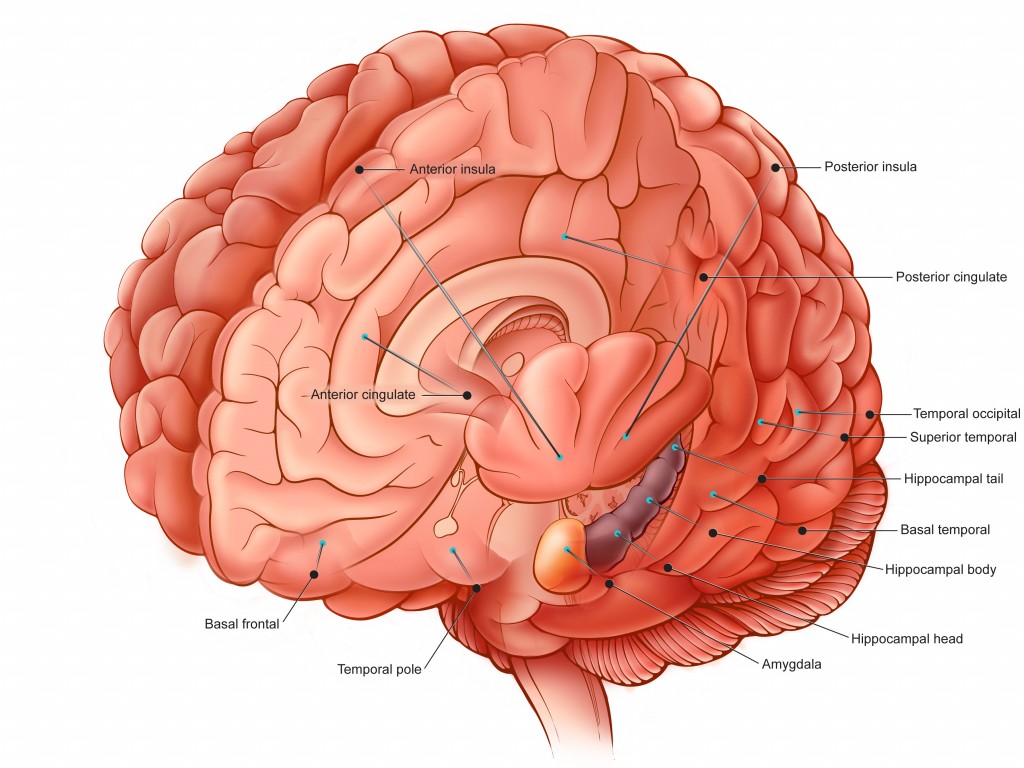
This illustration shows the locations for placing stereotaxic depth electrodes for electroencephalograph (EEG) monitoring. The purpose of the electrodes is to monitor a patient during a seizure in hopes of identifying the epileptogenic zone, to better treat the patient.
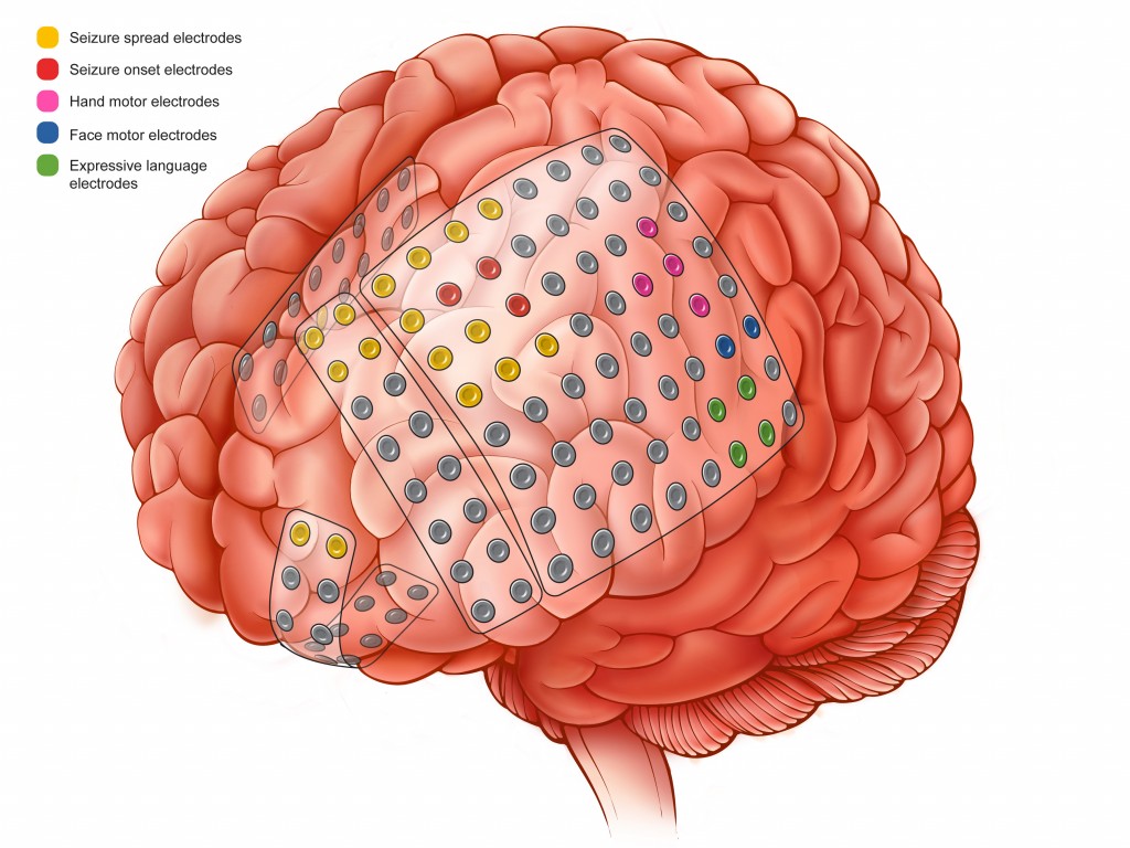
This illustration shows a different method for monitoring a patient during a seizure. A grid of subdural electrodes is placed intracranially. The purpose of the electrodes is to monitor a patient during a seizure in hopes of identifying the epileptogenic zone, to better treat the patient.